Suhasa Kodandaramaiah Awarded NIH Grant for Recording Neural Activity

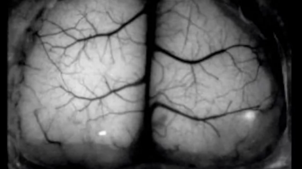
The NIH National Institute of Neurological Disorders and Stroke awarded $1.8 million to professors Suhasa Kodandaramaiah and Tim Ebner (Neuroscience) to develop better cranial exoskeletons for recording neural activity in mice. Their project, "Robot assisted brain-wide neural recordings and comprehensive behavioral monitoring in freely behaving mice," has the potential to reveal how brain-wide neural circuit computations are involved in everything from simple decision making to more complex cognitive tasks. Being able to better track and understand these computations will allow for better treatment of a wide variety of neurological diseases including Parkinson's, Alzheimers, and dementia — all diseases where such neural computations are disrupted.
Currently available technologies for recording neural activity in freely behaving animals suffer reduced performance from miniaturization. The team aims to engineer a cranial exoskeleton that attaches to the animal’s head and is capable of maneuvering neural recording hardware weighing orders of magnitude more than the animal subject, in 6 degrees of freedom in response to an animal’s movements. The cranial exoskeleton will reveal the mechanisms by which neural computations function normally to produce complex behaviors and how those computations go awry in neurological and neuropsychiatric diseases.
The project will be led by ME postdoc James Hope, working with graduate students Pin-Hao Cheng and Travis Beckerle, along with researcher Zoey Viavattine.
Drs. Ebner and Kodandaramaiah lead the Imaging Cells During Behavior core within the University of Minnesota's Center for Neural Circuits in Addiction. Their core is already assisting researchers at the UMN offering offers a range of imaging modalities to monitor brain activity in behaving animals across a range of spatial and temporal scales including: fiber photometry, head-mounted miniature microscopes (“miniscopes”) and novel wide field-of-view optical imaging during behavior at both the mesoscopic and cellular levels.