Ion Beam Analysis
The IBA lab was recently featured in a new "Did you know?" column of our CharFac newsletter. Read more on the "Did you know?" page.
Description
Ion Beam Analysis (IBA) uses a high-energy, light ion beam (typically He++, i.e., nuclei: alpha particles) to probe elemental composition as a function of depth (microns) with a depth resolution of 10-50 nm. In the core IBA method, Rutherford backscattering (RBS), the energy distribution of backscattered ions (He++) quantifies the depth distribution for a given element, while also dispersing the various elemental signals along the same energy scale. RBS is a fast, nondestructive (i.e., nonsputtering) and standardless techniques to quantify absolute atomic ratios (stoichiometry) in compounds or mixtures, insensitive to chemical environment. It can determine the film thickness given knowledge of atomic density, or density (thereby porosity) given knowledge of film thickness (e.g., from a SEM cross section image). In more advanced work one can gauge structural disorder in single crystals or epitaxial films via ion channeling methods.
Characteristic X-rays are also emitted from different target elements because of core electrons ejected by passing He++ nuclei. (Indeed it is these excitations, by taking kinetic energy away from the He++, that generate the perception of depth in RBS.) The X-ray emission spectrum (akin to EDS in SEM) can be used to ensure the accurate identification of similar mass elements (i.e., heavy impurities in a glass). Even higher-energy gamma rays emitted via nuclear reactions can provide excellent sensitivity for certain light elements such as Li and F. Finally, one can also tilt the sample and detect recoiling protons and deuterons from the sample, and thereby measure a hydrogen depth profile.
Thus Ion Beam Analysis is a broad term (worth wiki-ing or googling; other introductory literature can be provided) that involves several specific techniques with their own acronyms, namely:
- Rutherford backscattering spectrometry (RBS) to probe elements from Be on up through the heaviest elements in the periodic table
- Forward recoil spectrometry (FReS) or elastic recoil detection analysis (ERDA) of hydrogen (protons, deuterons)
- Nuclear reaction analysis (NRA) for enhanced sensitivity to elements in rows 1-3 of periodic table
- Particle induced gamma ray emission (PIGE) primarily for Li and F analysis
- Particle induced X-ray emission (PIXE) primarily for Na and heavier elements
- Ion channeling based crystallinity analysis for single crystal or epitaxial samples
Equipment
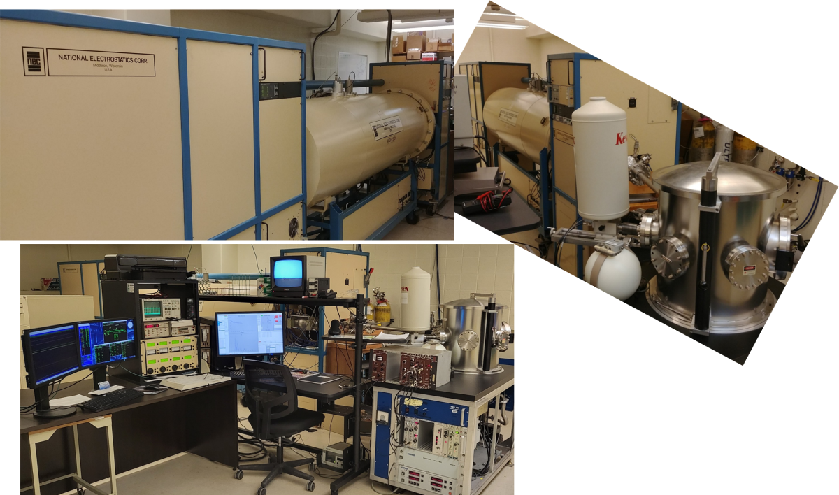
- MAS 1700 pelletron tandem ion accelerator (5SDH) equipped with charge exchange RF plasma source by National Electrostatics Corporation (NEC).
- Analytical end station (RBS 400) by Charles Evans & Associates, controlled by NEC DAQ PC.
Simultaneously operating detectors include
- Fixed ion detector at 165° for Rutherford backscattering spectrometry (RBS).
- (optional) Movable ion detector (90° - 150°) for grazing-angle scattering (enhanced surface sensitivity).
- Fixed ion detector at 30° for FReS
- Scintillation NaI(Tl) gamma-ray detector for PIGE
- Retractable Si(Li) X-ray detector for PIXE
Other elements of the control systems include
- single PC control (win10, added in 2021) of data acquisition and goniometer (translational and rotational movement of sample including algorithmic);
- simultaneous collection of RBS, FReS, PIGE, and PIXE spectra.
- automated sequential collection of data on multiple samples via defined queue of positions/tilts (beam located on luminescent reference sample, then drawn on CRT screen for sample positioning), one queue per beam energy;
- software/DAQ routines for finding channeling orientation of sample, plus DAQ while sweeping tilt through axial channel or spectral collection at channeled vs unchanneled orientations;
- manual selection of beam aperture in front of analytical chamber, from a range of shapes/sizes (default 2x2 mm, 1x1 mm also used quite frequently, but smaller options measured in hundreds of microns are also available);
- Linux computer and knobset control (“Accelnet” by NEC, added in 2017) of ion beam parameters such as energy, charge state, current, focusing;
Accessories
- Data analysis workstation (must log in but free for use) containing several data analysis programs: SimNRA, Quark, GUPIX and RUMP
- Custom batch data-file conversion from commercial DAQ software format to multicolumn text.
Applications and some specifications:
Rutherford backscattering spectrometry (RBS):
- Nondestructive, multi-elemental analysis
- Elemental composition (stoichiometry) without a standard (1 - 5% accuracy)
- Maximum depth of 2 - 20 microns
- Impurity distribution in depth
- Measure thickness (or density) of thin films if the film density (or thickness) is known.
- Diffusion depth profiles between interfaces up to a few microns below the surface.
- Channeling-RBS can be used on single crystal samples or epitaxial films to
- determine unit cell location of dopants (e.g., on lattice vs interstitial)
- depth profile defect/dopant distribution in single crystal samples or epitaxial films
- do either of the preceding specific to element (i.e., probe crystallinity in ways that X-ray diffraction cannot)
Forward recoil spectrometry (FReS):
- Nondestructively and simultaneously determine hydrogen (protons and deuterons) concentration and depth profile in polymers or other solids down to ~0.1%
- Depth resolution of 30-80 nm and maximum depth of ~1 micron
- Standard configuration utilizes 75 degree sample tilt and 12 micron mylar foil at detector (to block nuclei larger than deuterons); thus beam shape is effectively widened by nearly a factor of 4 (prepare samples accordingly) along one dimension
Nuclear reaction analysis (NRA; scattering via nuclear absorption rather than coulomb repulsion, whether re-emitting same He++ nucleus or a proton/gamma ray):
- Nondestructively probe light elements (such as Li, B, C, O, N, F) with greater sensitivity.
- Unlike nonreactive techniques, NRA is isotopically sensitive.
- Channeling-NRA can be used.
Particle induced X-ray emission (PIXE):
- Nondestructive and multi-elemental analysis of trace elements with an excellent detection (down to sub ppm for certain elements).
- Used together with RBS for accurate mass identification of medium to heavy elements with similar masses.
- Elemental composition of magnetic films in which RBS does not have an enough mass resolution to resolve Mn-Fe-Co-Ni elements.
- Channeling-PIXE can be used.
Ion channeling analysis
- Assess crystallinity of MBE-grown thin films such as type of defect structures, impurity location in unit cell, type of atomic site, lattice strain and alignment in epitaxial growth.
- Channeling provides enhanced sensitivity to light elements in film when atop heavier single-crystal substrate.
More detailed specifications:
Ion beam:
- Accelerator terminal voltage tunable over 0.5-1.7 MV
- He+ and He++ beams in standard configuration with maximum energies of 3.4 and 5.1 MeV, respectively.
- Beam current on target up to a few tens of nanoamps depending on ion species and energies.
Particle detectors:
- Ortec Ultra ion detectors
- Kevex Retractable Si(Li) X-ray detector with 5 mm Be-window
- Canberra 2" x 2" NaI(Tl) gamma detector
Goniometer:
- Sample lateral movements: ± 25mm with a minimum step size of 0.001mm.
- Tilting movements: ± 90° around vertical axis and ± 20° around horizontal axis with a minimum step of 0.01°.
Sample construct:
- Can load as many samples as will fit in 2x2 inch (carbon taped onto stainless steel block)
- Typical sample size: 5 x 5 mm2 or 10 x 10 mm2 for RBS/NRA/PIXE/Channeling and 5 x 15 mm2 for FReS. Minimum size 1 x 1 mm2, maximum size 50 x 50 mm2 (i.e., requiring entire block)
- Overall sample thickness is typically no more than 5 mm, but thicker samples can be accommodated with special preparation (cannot displace block along Y in this case, only X, thus far fewer samples can be accommodated in a single venting).
- Sample has to be a vacuum-compatible solid with reasonably smooth surface.
Sample Data
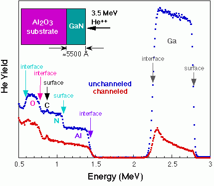
The above compares data taken at random sample orientation (unchanneled) and under axial channeling conditions. The colored arrows denote the energies of backscattered He nuclei from different elements at two depth locations. A 3.5 MeV beam energy enables certain nuclear reactions, thereby increasing the sensitivity to N and C.
References
Handbook of modern ion beam materials analysis. Yongqiang Wang, Michael Anthony Nastasi. Materials Research Society (2009, 2nd ed.). ISBN 978-1-60511-217-6. OCLC 672203193.
"Thin film depth profiling by ion beam analysis", Chris Jeynes and Julien L. Colaux, Analyst 141, 5944–5985 (2016)
"Strong band gap reduction in highly mismatched alloy InAlBiAs grown by molecular beam epitaxy", Jing Zhang, Yuejing Wang; Shoaib Khalid; Anderson Janotti, Greg Haugstad, Joshua M. O. Zide,
J. Appl. Phys. 126, 095704 (2019).
Recent external clients of the IBA lab:
US Research Universities (public and private)
- Brown University
- Columbia University
- Cornell University
- Iowa State University
- North Dakota State University
- Pennsylvania State University
- Stanford University
- University of California - Berkeley
- University of California - Santa Barbara
- University of Colorado - Boulder
- University of Delaware
- University of Illinois - Urbana-Champaign
- University of Pennsylvania
- University of Wisconsin - Madison
- Washington University - Saint Louis
Minnesota (primarily) Undergraduate Institutions
- Augsburg College
- Carleton College
- Macalester College
- University of St. Thomas
Minnesota Industry / Commercial
- Abbott Laboratories
- Advanced Research Corporation
- Agnitron Technology
- Boston Scientific, Inc.
- Carestream Health, Inc.
- Committee Films (for History 2 television channel)
- Det-Tronics
- NVE Corporation
- Physical Electronics, Inc.
- Sage Electrochromics
- SVT Associates, Inc.
Other US Industry
- Dow Corning Corporation
Non-US Institutions
- CINVESTAV-IPN, Mexico