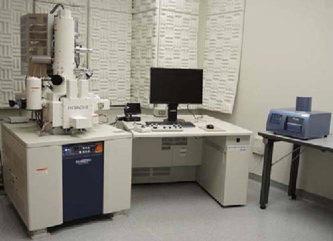
Hitachi SU8230 Field Emission Gun
Description:
Hitachi SU 8230 is a Field Emission Gun Scanning Electron Microscope (FEGSEM) capable of 1.1 nm resolution at 1.5 mm working distance at 1.0 kV landing voltage, and 0.8 nm resolution at 15 kV at 4 .0 mm working distance.
The newly designed Cold Field Emission (CFE) gun: complements the inherent high resolution and brightness of conventional CFE with increased probe current and beam stability. The source brightness (A / cm2/sr) exceeds that of any prior Hitachi SEM. Landing voltage range is 30 kV to 0.01 kV. (Beam Deceleration Mode; variable retarding bias, provides landing voltage range from 0.01 to 2kV (0.01 - 1 kV in 10V steps; 1.0 - 2.0 kV in 100V steps)
Magnification: ranges from 100X to 1,000,000X in High Magnification Mode; 20X-2000X in Low Magnification Mode. (Working distance dependent)
Electron detectors include:
- In-column Upper ExB detector with lens-integrated control electrode to provide either pure secondary (SE) or variable percentages of SE and inelastically scattered electrons (BSE)
- An in-column Top detector which receives high-angle elastically scattered electrons (BSE) for high resolution atomic number contrast at short working distances
- A chamber mounted Everhart-Thornley detector
- A 4+1 segment retractable below-lens semiconductor type BSE detector.
Sample chamber: 6 inch; includes an integrated LN2 cryo trap.
Stage: 5-axis motorized stage.
- Non-Cryo Mode: X/Y movement is 0-110 mm; Z: 1.5 - 40 mm; Tilt: -5 to +70; Rotation: 360.
- Cryo Mode: X/Y movement is ~ +/- 9 mm; Z: 1.5 - 40 mm; Tilt: 30 at Z=8mm or greater; 15 at 5 mm; 0 at 1.5 mm. Rotation: 0 (Locked!)
Vacuum system: Specimen chamber; Turbo + scroll dry pump. Gun / column; Ion pumps X 3, + Non- Evaporative Getter (NEG).
Charge Suppression Scan: imaging mode helps reduce charge artifact.
Applications:
- High-resolution topographic contrast (Se) and atomic number contrast (BSe) imaging of biological and non-biological samples in both room temperature and cryo modes.
- Ultra-low landing voltage imaging of beam sensitive or charge prone samples.
- Particularly applicable to imaging of immunogold labeled individual cells and bacteria, analysis of semiconductor thin films, polymers, hydrogels, etc.
Specifications:
- High-resolution topographic contrast (Se) and atomic number contrast (BSe) imaging of biological and non-biological samples in both room temperature and cryo modes
- Cold field-emission gun with "Mild Flashing" technology.
- 6 inch sample chamber; integrated LN2 cryo trap.
- Resolution: 0.8 nm resolution at 15 kV at 4.0 mm WD; 1.1 nm resolution at 1.0 kV landing voltage at 1.5 mm WD.
- Magnification ranges from 20 X to 1,000,000X. (See Description)
- In-column Upper ExB detector; In-column Top Detector, In-Chamber lower ET detector; 4+1 segment semiconductor (PD-BSE) detector. (See Description)
- Leica VCT-500 Cryo Stage / Cryo Transfer System
- Thermo Noran System 7 EDS (See Description)
Equipment/Accessories:
- Leica VCT500 Cryo Stage / Cryo Transfer System
- Thermo Noran System 7 Spectral Imaging (EDS) System with UltraDry Si Drift Detector
Capabilities:
- Sample Size, Non-cryo: ~ 150 mm Diameter
- Sample Size, Cryo: ~ 18 mm X 18 mm