Scientists use unique combination of techniques to study synapse formation and modifications in vivo

Study has implications for our understanding of learning, memory formation, and the overall plasticity of the brain
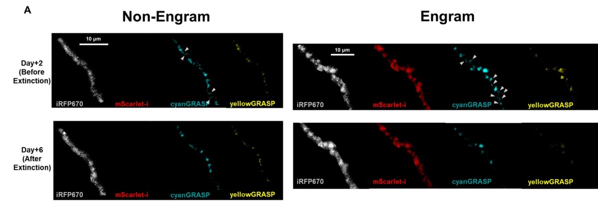
Research carried out by scientists from the University of Minnesota Twin Cities and Seoul National University in Korea has found an increase in the synaptic population among engram dendrites after memory formation, and a noticeable decrease in the synaptic population after memory extinction in live mice. A combination of techniques adopted by the team allowed them to gather longitudinal data on synaptic dynamics in vivo, which is a first in studies of this nature. The technique as well as the results of the study have implications for furthering the understanding of learning, memory formation and extinction, and the overall plasticity of the brain. The study was jointly conducted by ECE’s Professor Hye Yoon Park with Professor Bong-Kiun Kaang of Seoul National University, and their teams, and is published in Current Biology in a paper titled, “Hippocampal engram networks for fear memory recruit new synapses and modify pre-existing synapses in vivo.”
Engram cells, the cells that encode memories, are aided in the task of memory creation by structures called synapse. A synapse, the connection between cells or neurons, functions as a site for the transmission of messages or signals between cells: nerve impulses are sent from the axon (the long thin cable like structure that carries signals away from a neuron) of a sending neuron to the dendrite (the tree like projections on a neuron) of a receiving neuron.
The research conducted by the team is particularly significant for the insights it provides on the nature of the dynamics of the synapses through contextual fear memory formation, and fear memory extinction.
Previously, technical constraints attributed to the use of the dual-eGRASP system alone limited studies to gathering data from subjects at a specific memory state. However, the combination of two-photon imaging with the dual-eGRASP system overcomes the challenges posed by the use of the dual-eGRASP (enhanced green fluorescent protein reconstitution across synaptic partners) system alone. The newly developed combination of methods has dramatically changed the level of access, and the team was able to track the same synapse on specific dendrites over an extended period of time in vivo. This allowed the researchers to gather longitudinal data on the changing synaptic dynamics as it occurred across different memory states, memory formation and memory extinction.
Commenting on the significance of the techniques used in the study, Professor Park says, “Previously, researchers were able to detect these synapses in mice only after they sacrificed the mouse, which made it difficult to track those synapses over time. But now, we’ve made it possible to image the synapses in a live mouse brain over several days, so we can better understand what happens to them long-term. It’s the first time this has been done in a live mouse brain, so that’s pretty exciting news in this field.”
Study highlights
The key highlights of the study includes information surrounding the formation, location, and elimination of new synapses during memory formation and extinction. Firstly, the scientists noticed that there was a higher proportion of new synapses that formed in engram (E) dendrites as compared to non-engram (N) dendrites which point to a greater role the former may be playing in memory formation.
Secondly, just as memory formation correlated with the formation of new synapses, the extinction of memory resulted in reduced synapses. Interestingly, when the mice underwent fear extinction training it was the number of engram-engram (E-E) synapses that significantly reduced, but there was no such significant reduction in the E-N synapses.
Thirdly, researchers made some interesting findings regarding the distribution patterns of the newly formed synapses. They observed that although there were newly formed E-E and N-E synapses after memory formation, the distance between the new E-E synapses was less than the distance between other random synapses, and they tended to pair with dispersed existing E-E synapses rather than existing N-E synapses. This indicates that new synapses tend to form near pre-existing synapses, which results in the clustering of E-E synapses. They also tend to locate in dendritic areas that have greater capacity i.e., areas that are less crowded.
The research findings are significant to our understanding of learning, memory, and the plasticity of the brain. The study brings critical advancements to previous findings on engram cells and synaptic dynamics. Based on their findings, the researchers conclude that memory formation is an outcome of new as well as modified existing synapses, and propose that the new synapses induce modifications to the existing synapses which results in an expanded engram memory network. The clustering of synaptic engrams after a learning event have led the researchers to conclude that learning induces the clustering of synaptic engrams. Regarding fear memory extinction, the scientists point that data acquired during the study indicating the disappearance of some E-E synapses after fear memory extinction support the unlearning hypothesis of fear memory extinction.
The advances presented by the scientists are particularly significant; the combination of techniques has allowed them to differentiate between synapses that existed before the formation of fear memory and those that were created after the event. Similarly, they could also identify specific changes in the synaptic density after fear extinction. While the study does have its limitations, the results could lead to a deeper understanding of synaptic dynamics and synaptic clustering, their role in memory formation and learning, and where these results can be replicated in other regions of the brain.
Professor Bong-Kiun Kaang emphasizes the significance of the team’s research: “This work provides conclusive in vivo evidence for interregional synaptic engrams as the physical substrates of fear memory traces.”
The research was supported by the National Honor Scientist Program of Korea. Authors Byung Hun Lee, Jae Youn Shim, and Hye Yoon Park were supported by the Samsung Science and Technology Foundation and the National Research Foundation of Korea.
In addition to Park, the research team included Chaery Lee, Hyunsu Jung, Yongmin Sung, Hyopil Kim, Jooyoung Kim, Ji-il Kim, Dong Il Choi, and Bong-Kiun Kaang (Department of Biological Sciences, Seoul National University); Byung Hun Lee, and Jae Youn Shim (Department of Physics and Astronomy, Seoul National University); Chiwoo Lee (Interdisciplinary Program in Neuroscience, Seoul National University).
Read the full paper entitled, “Hippocampal engram networks for fear memory recruit new synapses and modify pre-existing synapses in vivo” in Current Biology, a peer-reviewed journal that publishes original research across all areas of biology.