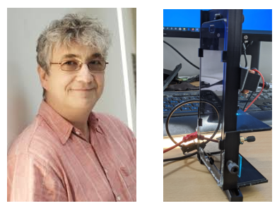
Physics Professor creating a smartphone device for COVID-19 diagnostic test

Professor Jochen Mueller of the School of Physics and Astronomy is part of a collaboration at the University of Minnesota that is building a device for smartphones that will help health care providers identify the COVID-19 virus, i.e., SARS-CoV-2. The collaboration is accelerating research during this unprecedented crisis directed at the development of a point-of-care diagnostic test for use by healthcare providers - particularly in resource poor locations - to rapidly identify the virus in under ten minutes.
Using funding from the UMN Medical School’s Rapid Response Grants Program, Mueller and Prof. Louis Mansky, Director of the Institute for Molecular Virology at the University of Minnesota have been working on the device around the clock for the past few weeks in order to create a functional prototype as soon as possible.
Mueller and Mansky are keeping the required supplies and reagents to a minimum, in order to help facilitate the mass production of test devices beyond the prototype. This is particularly important, as one of the critical limitations with current tests are limitations related to supply chain shortages. Sensitive and accurate testing that minimizes the likelihood of false positives and false negatives is essential for a test to meaningfully contribute to the urgent need for testing that will help allow for safely lifting stay-at-home restrictions and to identify those who may later become exposed to the virus.
Smartphone as microscope
Most smartphones have a built-in camera with an image sensor of high quality. By adding an external lens, the smartphone can be reconfigured to work as a microscope. The prototype created by Prof. Mueller’s research team uses a 3-D printed bezel to attach an inexpensive lens from a webcam to the back of a smartphone. A diode laser is incorporated into the 3-D print for detecting the virus by fluorescence, as well as a color filter to help improve the signal-to-noise ratio.
One of the challenges facing Mueller and Mansky has been accurate and sensitive virus detection. Mueller says that proteins on the surface of the virus particle are good targets for rapid virus detection. The SARS-CoV-2 spike protein is critical for virus entry into cells, and many copies of this protein decorate the surface of the virus particle, which visually creates a crown (i.e., corona) on the surface of the virus that can be seen by transmission electron microscopy. The SARS-CoV-2 spike protein interacts with the angiotensin converting enzyme 2 receptor protein, which is located on the surface of many cell types in the body, including the oral and nasal mucosa, nasopharynx, and lung.
Mansky and his research team are validating a virus detection assay that targets the SARS-CoV-2 spike protein using a fluorescently-labeled probe. The fluorescent label serves as the optical readout for the presence of the virus by the smartphone camera. Postdoc Rayna Addabbo is continuing this validation process in Dr. Mueller’s lab by using non-infectious virus particles carrying the spike protein. John Kohler, a Ph.D. student in Mueller’s group, is using state-of-the-art fluorescence microscopy to help validate the sensitivity of the smartphone-based imaging device.
Fundamental research
As a novel coronavirus, Mueller and Mansky also seek to better understand SARS-CoV-2 replication and particle assembly in order to identify therapeutic targets for novel intervention strategies. They collaborate with Prof. Wei Zhang, an expert in cryo-electron microscopy, who oversees cryo-electron microscopy at the UMN as part of her role in the Characterization Facility.