BME research develops novel approaches to identify targets for atrial fibrillation ablation
March 3, 2021 — A team of researchers from the University of Minnesota (Dr. Henri Roukoz, Vasanth Ravikumar) and Mayo Clinic (Dr. Siva Mulpuru), led by Prof. Alena Talkachova, is interested in developing new approaches to identify potential drivers for atrial fibrillation (AF) that could potentially help guide catheter ablation, a procedure for treating AF.
Complex cardiac arrhythmias, such as AF, have posed many challenges to physicians. Currently, in the US alone, it is estimated that 2.7-6.1 million people have AF. One treatment option is catheter ablation which targets the tissue responsible for the arrhythmia. However, AF recurrence rates remain high in part due to inaccurate localization of the sources of the arrhythmia, leading to additional health care costs and possible surgery.
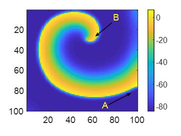
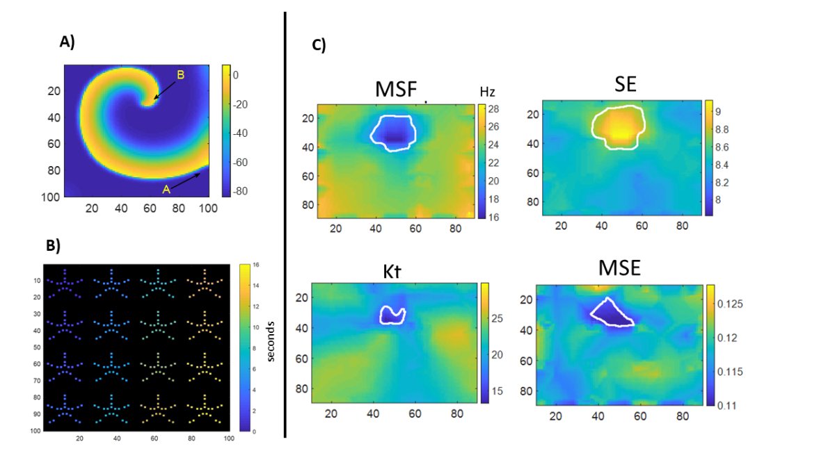
A team of researchers led by Prof. Alena Talkachova recently developed several frequency-based, information-based and statistic-based approaches that can be used for analysis of electrograms recorded during AF. In the manuscript, they evaluate the efficacy of these techniques to more accurately identify AF drivers using simulated bipolar and unipolar electrograms under different clinical scenarios like the presence of noise, prior scar, reduced spatial resolution and sequential signal acquisition.
This could fill a gap in current electro-anatomical mapping systems and more specifically provide a personalized therapy to map AF substrates on a patient-basis to improve ablation outcomes and to reduce AF recurrence rates.
Ravikumar V, Annoni E, Parthiban P, Zlochiver S, Roukoz H, Mulpuru S, and Tolkacheva E.G, 'Novel mapping techniques for rotor core detection using simulated intracardiac electrograms.' J Cardiovasc Electrophysiol. 2021;1–13. https://doi.org/10.1111/jce.14948