Biomedical optics & imaging
Imaging tools to better understand the brain
The Akkin Lab develops non-contact optical imaging tools to study tissue structure and function, with an emphasis on better understanding the brain. Non-invasive or minimally invasive applications in medicine are possible, because the techniques use back-scattered light.

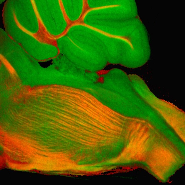
Imaging brain-body communication
Taylor Cannon’s lab is developing new optical imaging methods to capture how disruptions in central and peripheral nervous system activity drive disorder and disease. Visualizing cell signaling deep in the body paves the way for new clinical insights, diagnostics, and therapies.
Engineering the immune response
The Hartwell Immunoengineering Lab uses biomolecular engineering, drug delivery, and immunology to develop molecular vaccines and immunotherapies that direct the immune response towards activation or tolerance by targeting specific cells and tissues, with a focus on the mucosal immune system.
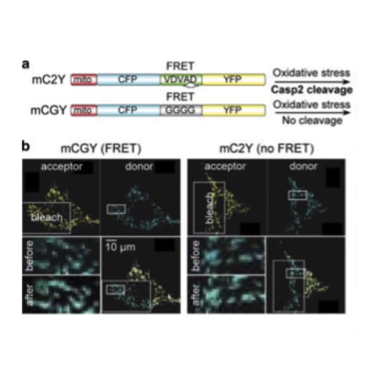
Aging and neurodegenerative diseases
Aging is the major risk for neurodegenerative diseases (NDs). Dr Herman and his colleagues have elucidated the role the Caspase-2 plays in neurodegenerative diseases. Current efforts are centered on the regulation of Caspase-2 mediated proteolysis of tau in NDs.
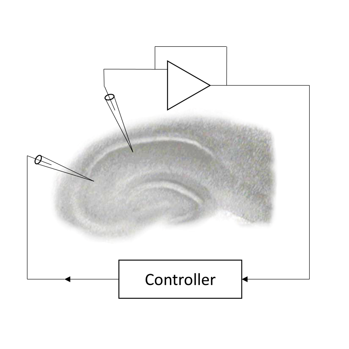
Optimizing stimulation therapy algorithms
The NeuralNetoff lab aims to give patients the best outcomes from electrical stimulation, a patient-specific therapy that varies widely in its effectiveness. They’re testing therapies and optimizing algorithms, to help people with conditions like Parkinson’s, epilepsy, and chronic pain.

Technology that tackles brain disorders
Alexander Opitz's lab aims to improve non-invasive brain stimulation technology, which people respond very differently to. The team is identifying individual predictors, to help create a future where there are personalized treatments for brain disorders like depression.
Bioengineering cancer therapies
Paolo Provenzano’s lab is developing new ways to combat cancer. Approaches include re-engineering tumor microenvironments to remove tumor-promoting cues, enhancing drug delivery, promoting anti-tumor immune responses, and developing next-generation cell-based therapies.
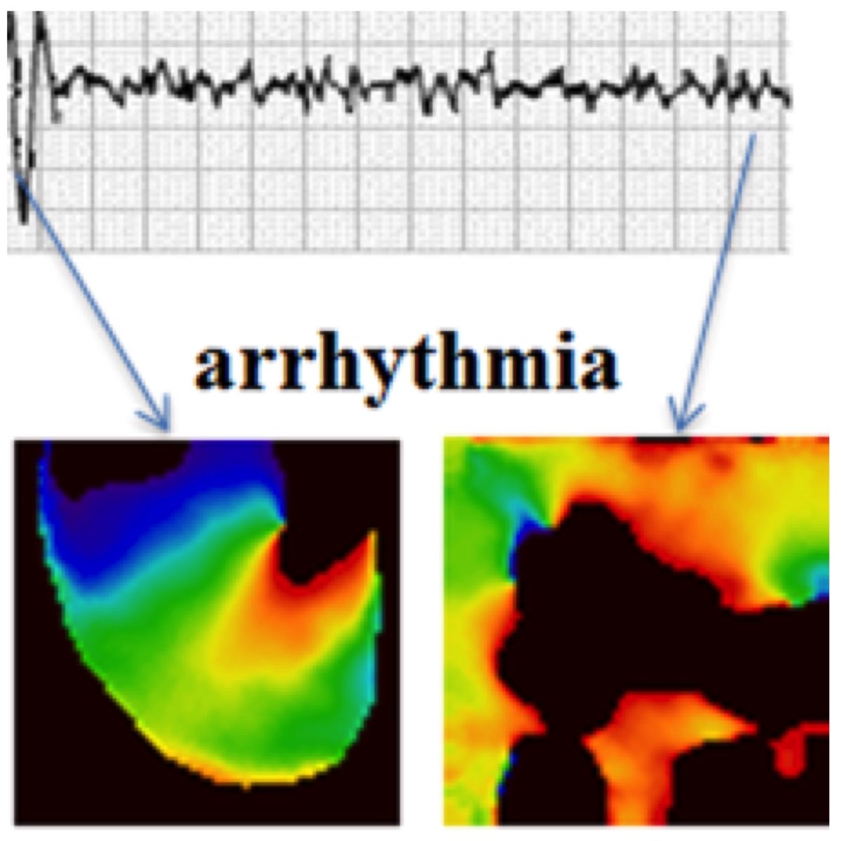
Prediction and prevention of cardiac arrhythmias
Alena Talkachova’s group visualizes electrical activity in the heart and small patches of cardiac tissue. They use nonlinear dynamics approaches to predict transition from normal to abnormal cardiac rhythms, and to prevent arrhythmias in the heart. They also develop novel tools to guide mapping-specific ablation in patients with atrial fibrillation.
Research from our graduate faculty

Technology for ultra high field magnetic resonance systems
The Adriany Lab designs designs novel magnetic resonance imaging array combinations for imaging at 7 Tesla (300 MHz), 10.5 Tesla (450 MHz), and 16.4 Tesla (700 MHz).

Quantitative imaging of brain energy metabolism
Wei Chen’s lab has developed a variety of X-nuclear magnetic resonance spectroscopic imaging methodologies and advanced radiofrequency coil technologies for noninvasively studying cellular metabolism, bioenergetics, function and dysfunction of the brain and other organs at ultrahigh field.

Geometry for cells
Meghan Driscoll’s lab aims to understand the functions of cell geometry and dynamics for cancer and immune cells. To do so, the lab combines advanced microscopy with the development of machine learning and computer graphics algorithms.

Advanced brain imaging for neuromodulation
The Harel Lab develops imaging tools that use ultra-high field MRI technology (e.g., 7 Tesla) to visualize the brain’s intricate anatomical structures. Research focuses on generating highly detailed maps of brain target areas and their anatomical connections — critical for improving deep brain stimulation treatments.

Quantitative MRI of musculoskeletal disorders
Casey Johnson's lab is developing quantitative MRI techniques to better understand, stage, and inform treatment of musculoskeletal disorders. Research in the lab includes the technical development of MRI methodologies and their validation in preclinical model and human studies.

Understanding brain-wide circuits mediating complex behaviors
Bridging neuroscience, genomics, and engineering, Suhasa Kodandaramaiah’s laboratory invents transformative technologies for ultra-large-scale neural recordings during complex cognitive behaviors. More recently, efforts have expanded to apply these tools to fundamental neuroscience questions.

Brain connectomics: From algorithms to applications
Diffusion magnetic resonance imaging is revolutionizing brain connectivity mapping, rapidly advancing our understanding of neurological and psychiatric illnesses. Christophe Lenglet's lab aims to create imaging and computational methods to uncover the mechanisms underlying these conditions and discover biomarkers that will accelerate clinical trials.

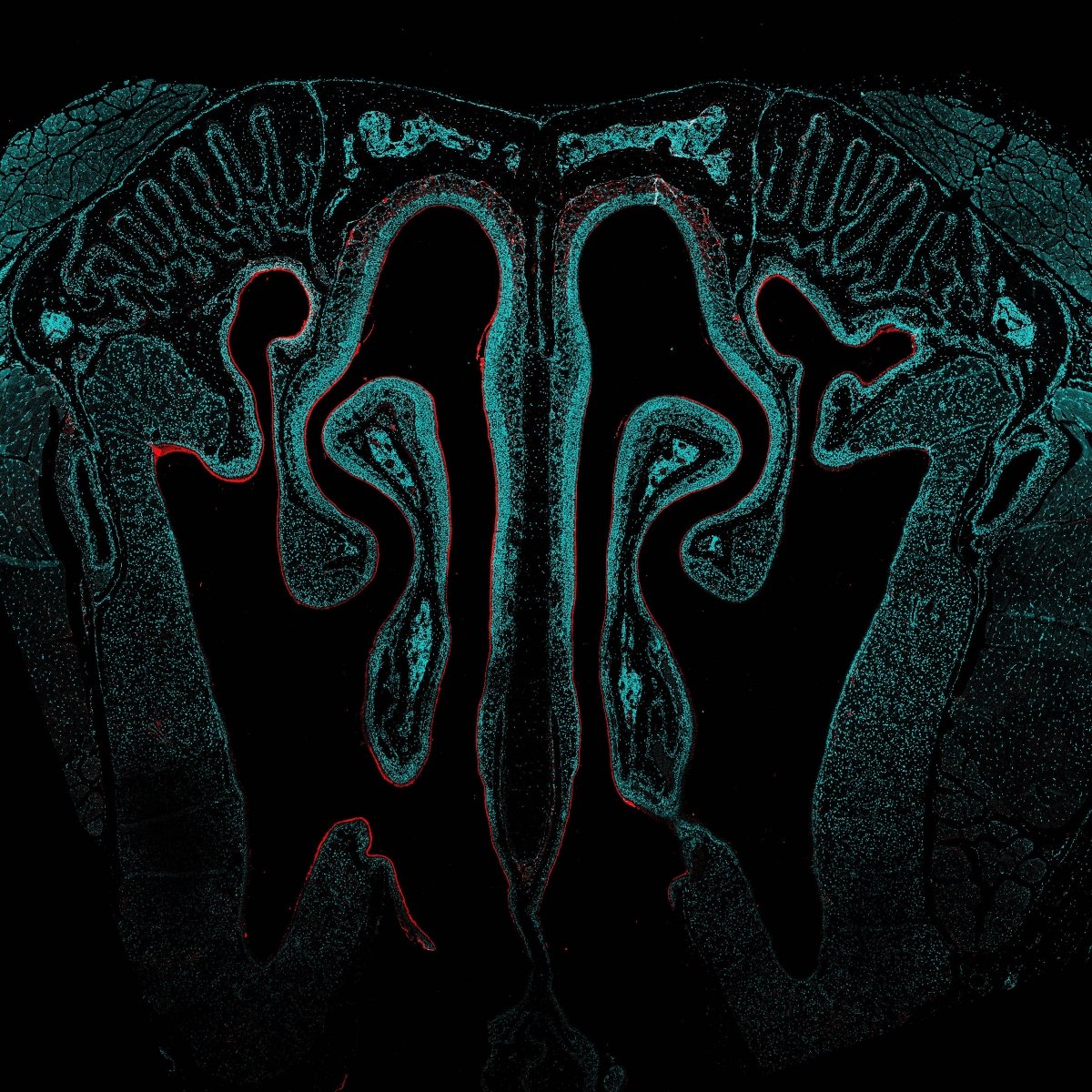
Quantitative virus and cancer imaging
Louis Mansky’s research group is addressing questions involving macromolecular assemblies important in virus and cancer pathobiology with quantitative fluorescence, electron imaging techniques, and large data informatics.

MRI technology development and translation
The Metzger Lab focuses on MRI technology development and its translation to in vivo systems. Research spans the breadth of MRI hardware development, electromagnetic simulations, quantitative imaging, predictive modeling, and developing RF management strategies to improve image quality and safety.

Tracking RNA in the living brain
Hye Yoon Park’s lab visualizes how neurons store memories by tracking RNA molecules in real time. This helps reveal how learning shapes the brain and opens new paths for diagnosing and treating neurological diseases.

Development of large-scale brain networks
Gordon Smith’s lab aims to understand how the large-scale networks that process sensory information and guide behavior form during development. His team uses cutting-edge optical tools together with computational modeling to measure and manipulate networks in vivo in the developing cortex.

Biophysical probes for cardiac drug discovery
The Thomas Lab uses molecular biophysics to create fluorescent probes for detecting protein structural changes, with the goal of discovering small-molecule drugs for treatment of disease, focused on muscle (cardiac and skeletal), but also including cancer.

Technologies to advance human brain MRI
Xiaoping Wu's laboratory focuses on advancing high-resolution brain MRI at ultrahigh magnetic field strengths. By developing data acquisition and image reconstruction techniques, the lab produces superior structural and functional brain images, unlocking new insights into brain anatomy, connectivity, and function.

Neural foundations of complex cognition
Jan Zimmermann’s lab explores the neural foundation of decision-making. The interdisciplinary team uses approaches from neuroscience, economics, psychology, math and physics to figure out how organisms adaptively use their finite neural coding capacity to make choices.