Biomolecular, cellular, & tissue engineering
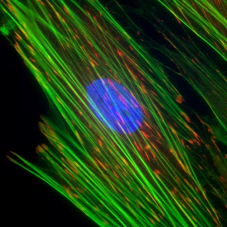
Cellular mechanics and mechanobiology
Many cells in tissues are exposed to dynamic mechanical perturbations, which require constant feedback by those cells to maintain tissue function. The Alford Lab uses novel microfabrication and computational methods to better understand this cellular adaptation in development and disease.
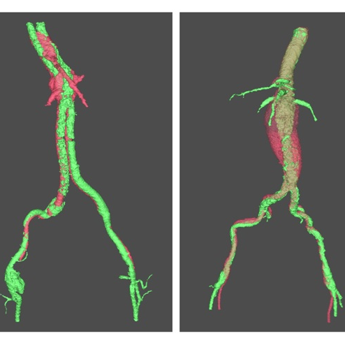
Tissue mechanics, aneurysms, and pain
The Barocas research group explores the relationship between tissue architecture and mechanics using multiscale computational models and mechanical experiments. Currently, they’re researching how aneurysms grow and fail, and how spinal load leads to injury or pain.
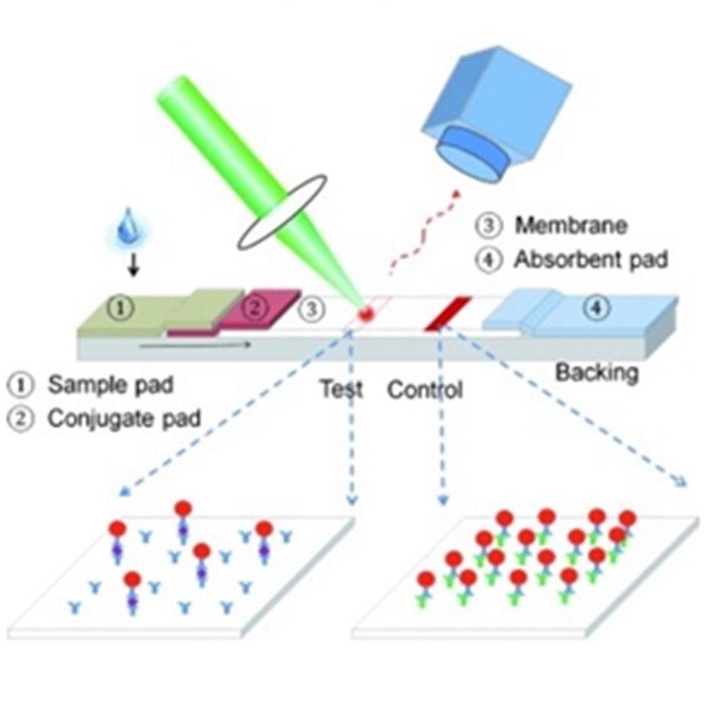
Thermally manipulating biomaterials
The Bischof group studies the thermophysical and biological changes within biomaterials after thermal manipulations. For example, they’re using nanoparticles to rewarm preserved tissues and organs and developing energy-based technologies to improve cancer immunotherapies.
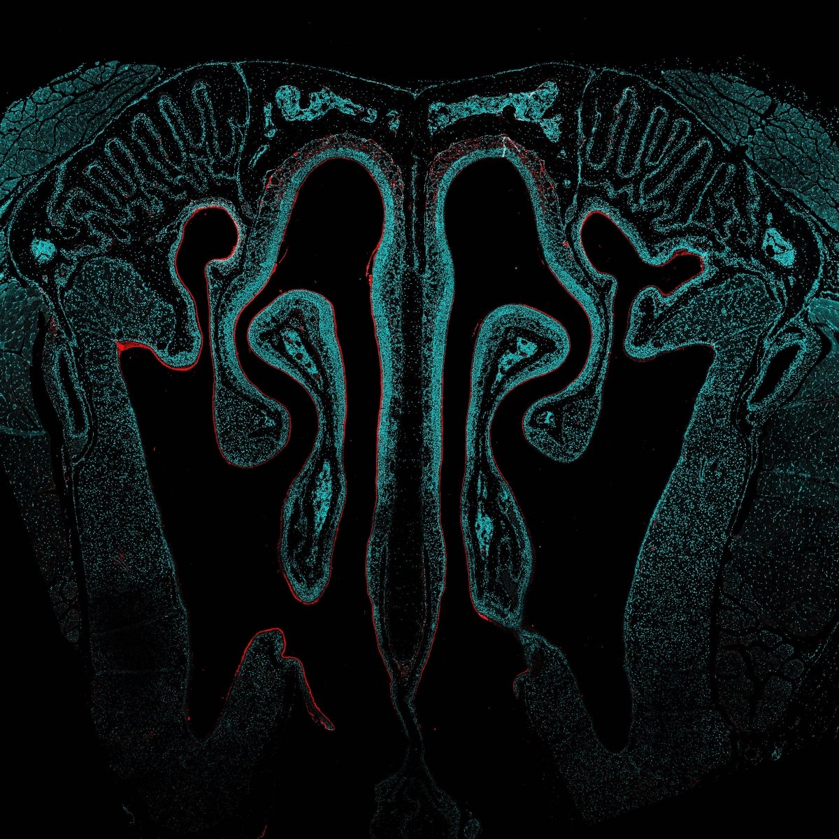
Engineering the immune response
The Hartwell Immunoengineering Lab uses biomolecular engineering, drug delivery, and immunology to develop molecular vaccines and immunotherapies that direct the immune response towards activation or tolerance by targeting specific cells and tissues, with a focus on the mucosal immune system.
Aging and neurodegenerative diseases
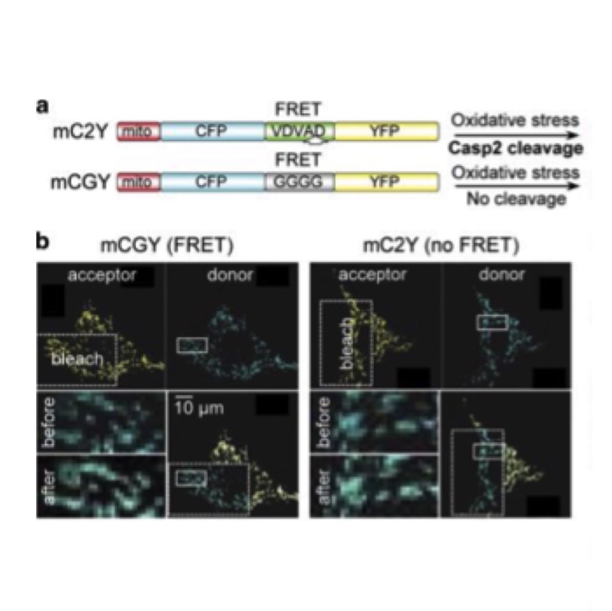
Aging is the major risk for neurodegenerative diseases (NDs). Dr Herman and his colleagues have elucidated the role the Caspase-2 plays in neurodegenerative diseases. Current efforts are centered on the regulation of Caspase-2 mediated proteolysis of tau in NDs.
How cellular functions go awry
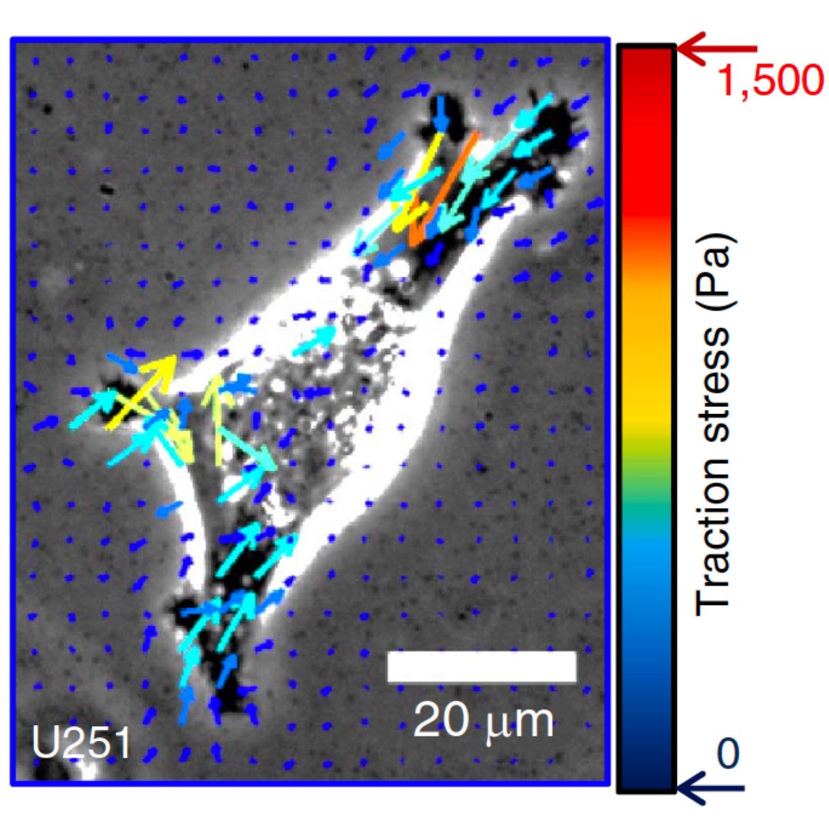
The Odde Lab aims to understand basic cellular functions in the context of diseases such as brain cancer and Alzheimer's. The team develops physics-based models that predict cell behavior, then use computer simulation and live cell imaging to identify potential therapeutic strategies.
Bioprinting cardiac tissues
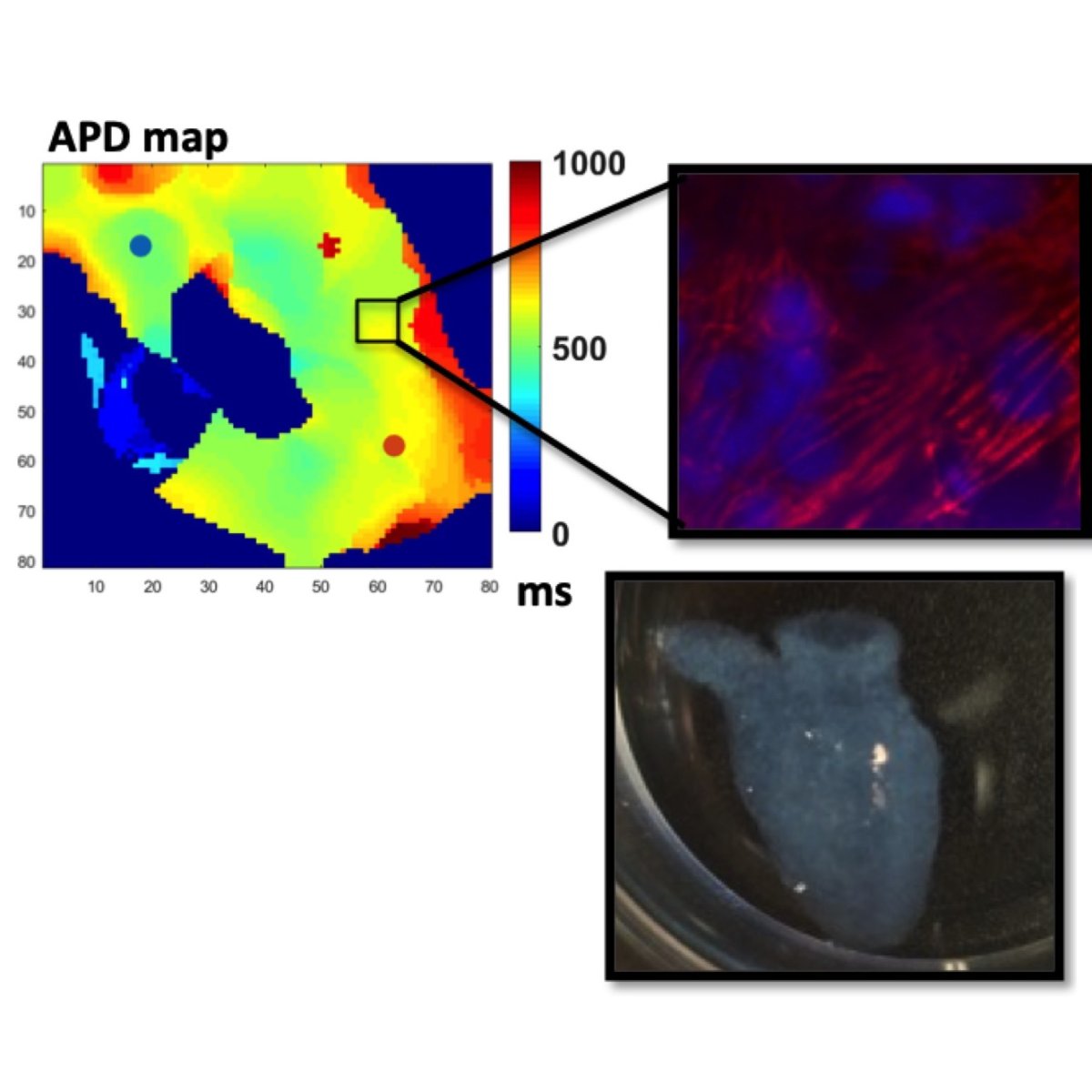
The Ogle Lab is pushing the boundaries of 3D cardiac bioprinting. They’ve created patches that can be adhered to failing hearts, which has successfully restored cardiac function in rodents. Plus, they’ve fabricated living hearts based on a human heart’s magnetic resonance imaging (MRI) data.
Bioengineering cancer therapies
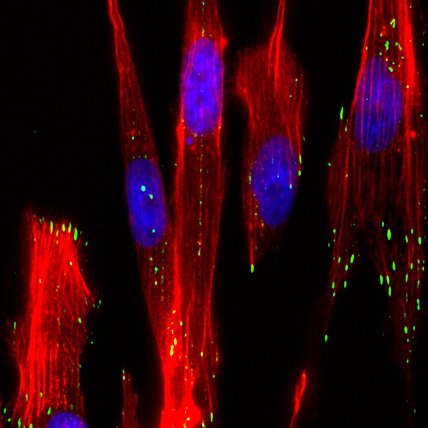
Paolo Provenzano’s lab is developing new ways to combat cancer. Approaches include re-engineering tumor microenvironments to remove tumor-promoting cues, enhancing drug delivery, promoting anti-tumor immune responses, and developing next-generation cell-based therapies.
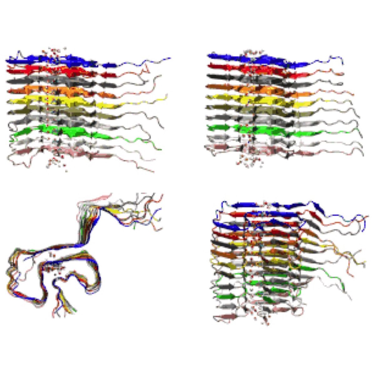
Discovering treatments at the molecular scale
The Sachs Lab is trying to explain how molecules malfunction in diseases like arthritis and Parkinson’s, to discover new treatment strategies. To do this, the team combines experimental biophysics, cell biology, and computational modeling using some of the world’s fastest supercomputers.
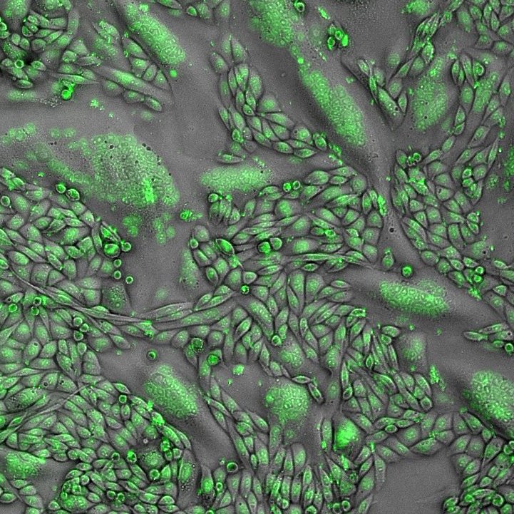
Understanding protein networks
The Sarkar laboratory uses approaches from biomolecular engineering and biology to better understand how protein networks drive health-related processes at the cellular level. Ultimately, this could lead to more effective therapeutics, such as to stop the proliferation of cancer.
![Shows 4 green-blue-black images taken with imaging technology. Includes red double arrows indicating nanogroove direction. Shows non-diseased (with zoomed in region), DMD [delta]ex52-54, and DMD[delta]ex31](/sites/cse.umn.edu/files/styles/folwell_full/public/shen-research-graphic-square.jpg.webp?itok=3JfoosZ0)
Engineering biomaterials to model diseases
Wei Shen’s laboratory engineers biomaterials, studies how they interact in microenvironments, and models diseases. They’ve created material for studying muscular dystrophy, a nanoparticle platform for antiviral therapy, and oxygen-releasing materials for cell-based therapy.
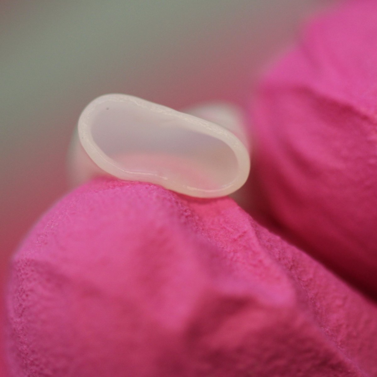
Living valves for growing bodies
Bob Tranquillo’s laboratory develops biologically engineered “off-the-shelf” vascular grafts, heart valves, and vein valves. They’ve shown the material, produced by skin cells, has the capacity to grow, which may transform the way pediatric congenital heart defects are treated.
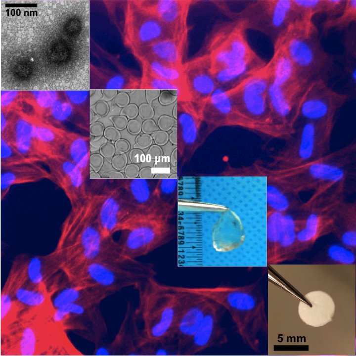
Polymers to deliver drugs, genes, and cells
Chun Wang’s laboratory develops polymeric materials to address unmet challenges in drug delivery. For example, they’re creating biodegradable polymers for cancer immunotherapies, including vaccines, and polymer wafers that’d be taken orally to deliver proteins and genes.
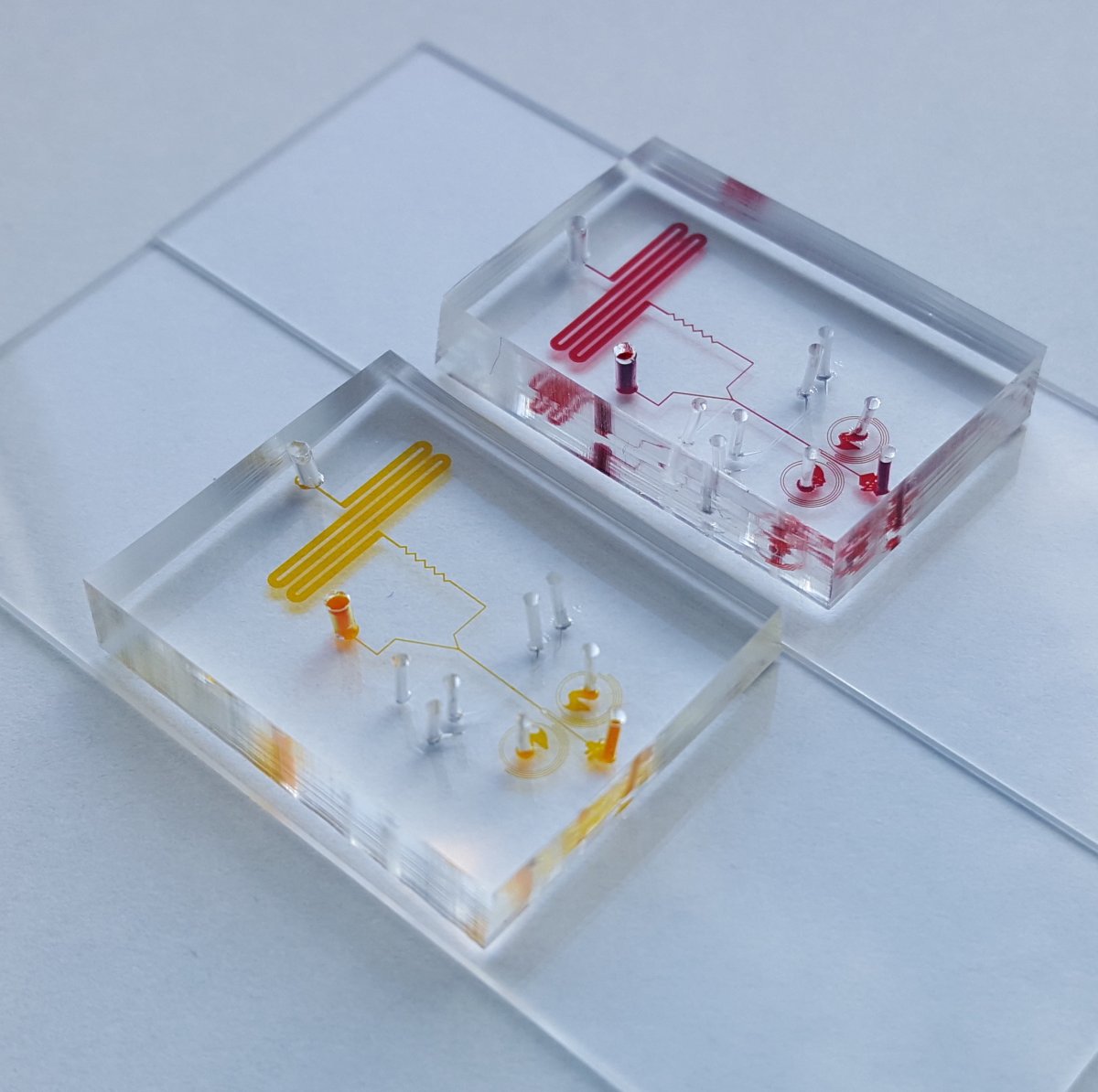
Microphysiologic systems to study disease
The Living Devices Lab is focused on building benchtop systems that mimic human disease outside the human body. We use microfluidics and microfabrication to create engineered tissues in which we control biological components and transport processes at the length scale that is relevant to physiology and pathology.

Pregnancy and soft tissue biomechanics
Kyoko Yoshida's lab studies how soft tissues grow and remodel to support a healthy pregnancy. They combine experimental and computational methods to uncover how mechanical and hormonal changes interact to drive dramatic tissue changes during pregnancy.