Cardiovascular engineering
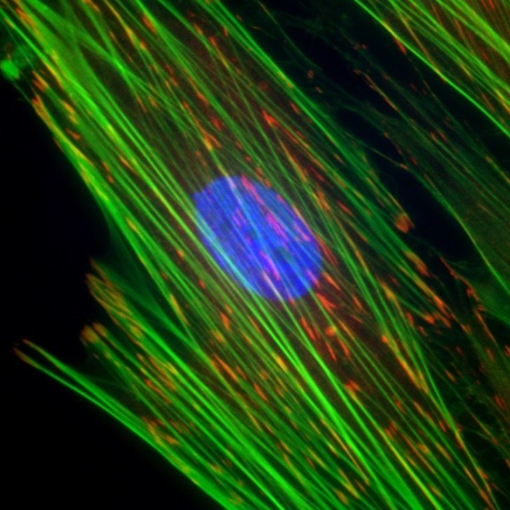
Cellular mechanics and mechanobiology
Many cells in tissues are exposed to dynamic mechanical perturbations, which require constant feedback by those cells to maintain tissue function. The Alford Lab uses novel microfabrication and computational methods to better understand this cellular adaptation in development and disease.
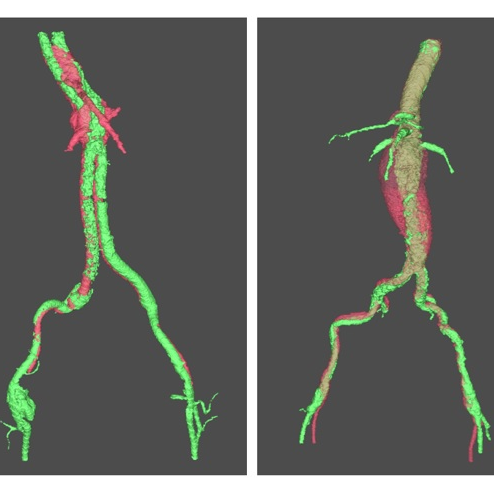
Tissue mechanics, aneurysms, and pain
The Barocas research group explores the relationship between tissue architecture and mechanics using multiscale computational models and mechanical experiments. Currently, they’re researching how aneurysms grow and fail, and how spinal load leads to injury or pain.
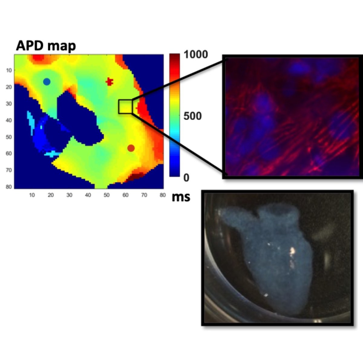
Bioprinting cardiac tissues
The Ogle Lab is pushing the boundaries of 3D cardiac bioprinting. They’ve created patches that can be adhered to failing hearts, which has successfully restored cardiac function in rodents. Plus, they’ve fabricated living hearts based on a human heart’s magnetic resonance imaging (MRI) data.
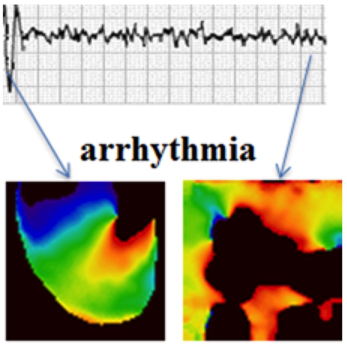
Prediction and prevention of cardiac arrhythmias
Alena Talkachova’s group visualizes electrical activity in the heart and small patches of cardiac tissue. They use nonlinear dynamics approaches to predict transition from normal to abnormal cardiac rhythms, and to prevent arrhythmias in the heart. They also develop novel tools to guide mapping-specific ablation in patients with atrial fibrillation.
Living valves for growing bodies
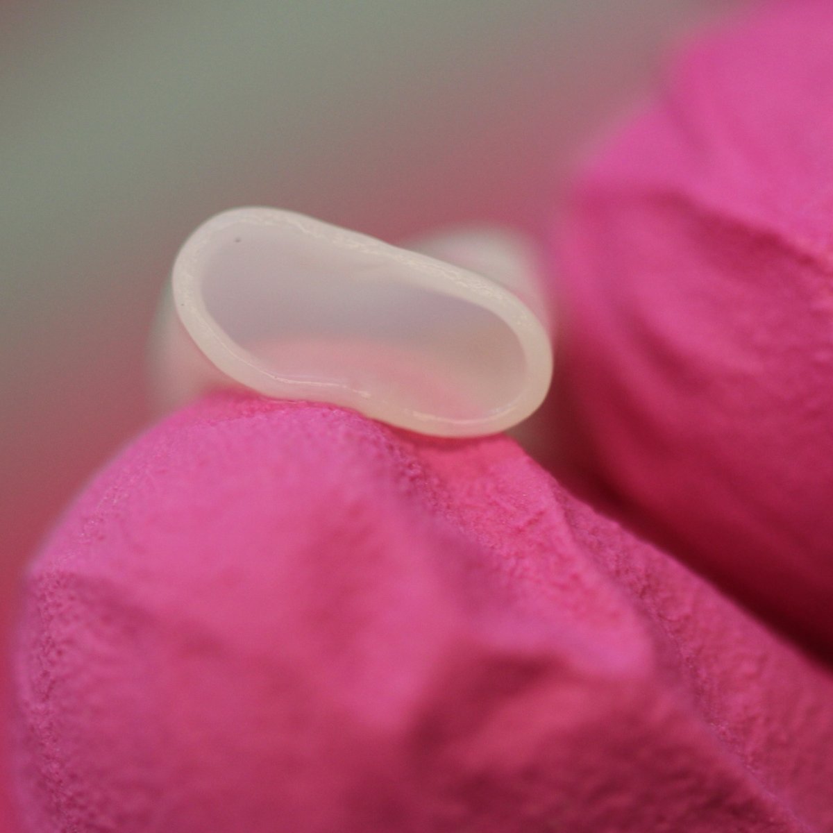
Bob Tranquillo’s laboratory develops biologically engineered “off-the-shelf” vascular grafts, heart valves, and vein valves. His team has shown the material, produced by skin cells, has the capacity to grow, which may transform the way pediatric congenital heart defects are treated.
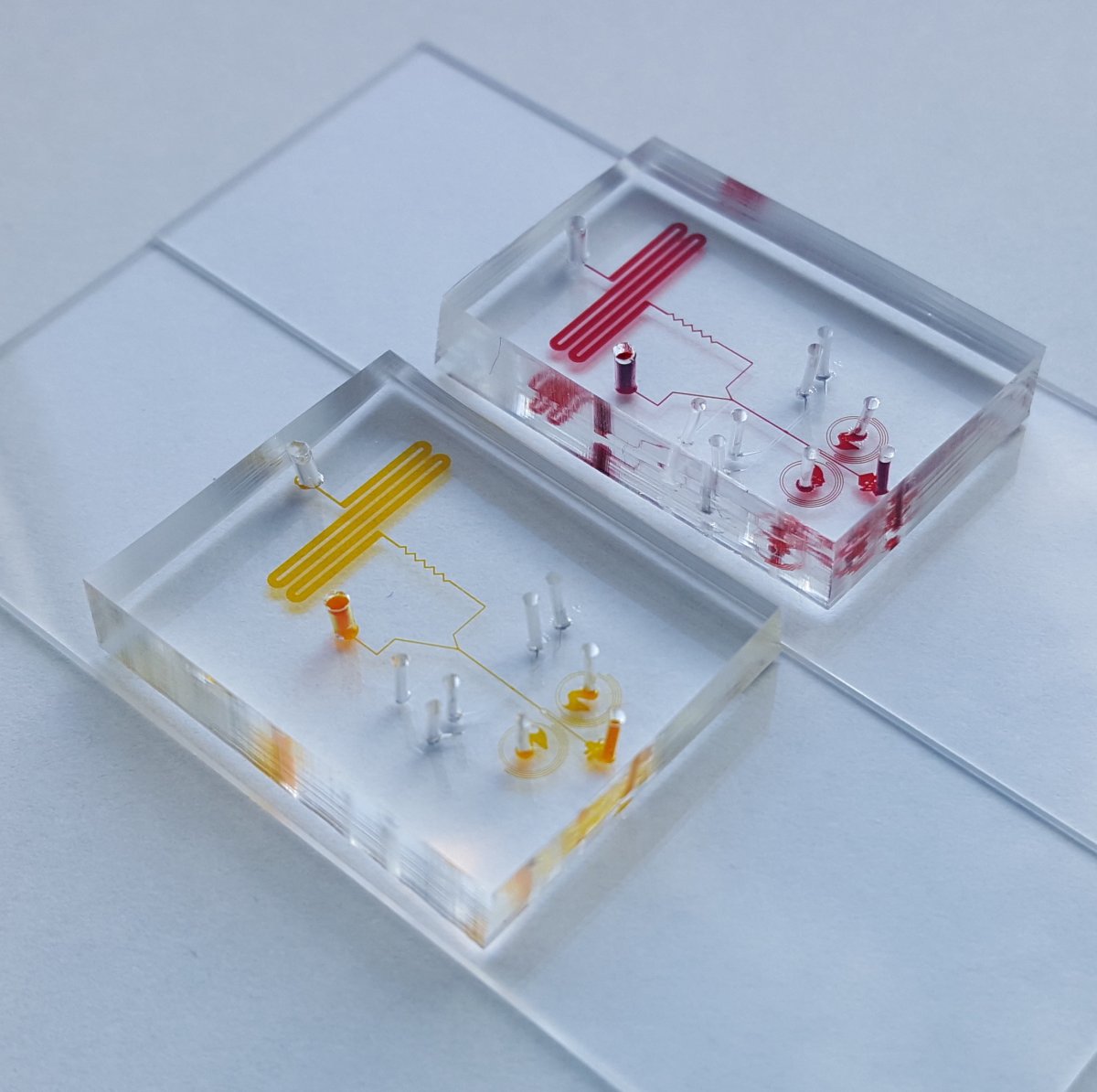
Microphysiologic systems to study disease
The Living Devices Lab is focused on building benchtop systems that mimic human disease outside the human body. We use microfluidics and microfabrication to create engineered tissues in which we control biological components and transport processes at the length scale that is relevant to physiology and pathology.

Pregnancy and soft tissue biomechanics
Kyoko Yoshida's lab studies how soft tissues grow and remodel to support a healthy pregnancy. They combine experimental and computational methods to uncover how mechanical and hormonal changes interact to drive dramatic tissue changes during pregnancy.
Research from our graduate faculty

Cardiac anatomy and physiology
The Visible Heart Laboratories focus on translational systems physiology, using multimodal imaging techniques of functional cardiac anatomies and device testing within large mammalian and human hearts.

Biophysical probes for cardiac drug discovery
The Thomas Lab uses molecular biophysics to create fluorescent probes for detecting protein structural changes, with the goal of discovering small-molecule drugs for treatment of disease, focused on muscle (cardiac and skeletal), but also including cancer.