Electron Microscopy

Electron microscopy, in its various forms and with myriad ancillary techniques, has become an indispensable characterization method for elucidating structure-function properties of nanoscale materials, molecular assemblies, and device architectures. The breadth of information that can be obtained is large and includes atomic-scale structure, electronic properties (e.g., density of states and plasmonic characteristics), and chemical composition (e.g., spatial element distribution, bond type, and oxidation state). Efforts in instrument development have led to significant advances in spatial, energy, and temporal resolutions; spherical-aberration correction enables formation of probe sizes well below 1 Å, monochromated electron guns can achieve energy resolutions below 100 meV, and pulsed-laser based methodologies that circumvent the millisecond resolution of digital detectors enable studies to be conducted on the femtosecond (i.e., 10-15 s) timescale. In situ and operando electron microscopy experiments – wherein materials are studied at elevated temperatures and pressures, immersed in liquids, and under electrical biasing or during mechanical deformation – are making possible the observation of atomic-scale morphological changes of catalyst particles in liquid phases, visualization of ferroelectric domain and domain wall dynamics, and the study of grain boundary and dislocation motion, to name a few. With its suite of microscopy capabilities, CEMS will continue to advance the field with innovative approaches to specimen preparation as well as via development of new techniques and instrumentation.

Atomic-resolution Imaging

electron 1b

With access to a variety of advanced scanning and transmission electron microscopes (TEMs) at UMN, including aberration-corrected scanning-TEM (STEM), almost all materials can be visualized and characterized at atomic-level precision. These capabilities allow us to study technologically important nanoscale materials and membranes, evaluate the growth quality of thin films, and understand structural issues often present in them. Different modes of imaging provide information about local atomic arrangement, composition, presence of stain, and much more. 

Related Faculty and Research Groups:

Ultrafast EM Imaging & Spectroscopy

electron 2

Application of femtosecond transmission electron microscopy (or ultrafast electron microscopy, UEM) and spectroscopy provide new ways of study phonon dynamics in the materials. We study defect-modulated phonon dynamics in bulk and layered materials and correlate coherent acoustic-phonon dynamics with structure and morphology of materials. Additionally, the effects of pulsed electron beams in the UTEM allows us better understanding specimen damage under highly controlled e-beam radiation. Further development and advancement of UEM are part of the activities in this area. 

Related Faculty and Research Groups:

Defects in Crystals

eleactron 3

In crystals, imperfections exist as various forms of defects covering a full dimensionality spectrum ranging from 0-D point defects, such as vacancies and dopants, to more extended defects, such as 1D dislocations and disclinations, 2-D grain boundaries and stacking faults and, last, 3-D voids and inclusions. Extended defects are particularly promising for applications because they run across the macroscopic crystal in certain directions and are atomically small in others. They can provide atomic-scale tunability of material properties.

Related Faculty and Research Groups:

Spectroscopy

electron 3

Aberration-corrected STEM, available at UMN, is equipped with a variety of analytical spectrometers allowing electron energy loss spectroscopy (EELS) for electronic structure measurements, and energy dispersive x-ray (EDX) spectroscopy for compositional analysis. Using core-level EELS, local electronic density of states specific for those atoms can be measured, and by using low-loss EELS, dielectric and optical properties are determined. This is vital for the in-depth characterization of technologically important nanoscale materials and devices.

Related Faculty and Research Groups:

Microscopy for Metals, Alloys & Ceramics

electron 5

There is a close relationship between the microstructure and mechanical behavior of materials. It is even more enhanced in nanoscale materials and materials in extreme environments such as high temperature, stresses, strain rates, and radiation. Electron microscopy is an excellent way to evaluate the atomic structure of materials and understand their mechanical behavior.   

Related Faculty and Research Groups:

Cryo-TEM & Cryo-SEM

electron 6

Cryogenic transmission and scanning electron microscopy (referred to as cryo-TEM and cryo-SEM) are used to understand and explain the nanostructure formation process steps, as well as the sensitivity to process variabilities in the formation of certain nanoparticles and long-lived nanoscale oil-in-water dispersions. 

Related Faculty and Research Groups:

Relevant Collaborative Partners and Core Facilities

Electron Microscopy research takes advantage of existing at the University of Minnesota-Twin Cities campus CSE Characterization Facility, which houses several cutting-edge STEMs, TEMs, SEMs, FIB, etc. along with sample preparation facility and dedicated staff. The faculty is located between Shepherd Lab (for hard materials) and in Nils Hasselmo Hall (for soft materials), both of which are just a short walk away from Amundson Hall. You can find the list of Electron Microscopes at CharFac website. UMN also provides excellent computing resources and services to support the computational side of our microscopy-based research. The Minnesota Supercomputing Institute, located in Walter Library, houses three high-performance computing clusters, providing more than 80,000 CPU and GPU cores to research groups at UMN, along with extensive cloud computing, data storage, and dedicated staff. 

Research in Electron Microscopy at CEMS is highly interdisciplinary. We collaborate with researchers from many departments across the university (Chemistry, Physics, ME, ECE, etc.) as well as research centers such as the UMN Materials Research Science and Engineering Center, and the UMN Minnesota Nano Center.

Major Funding Sources

Selected Publications

Expand all

One-dimensional intergrowths in two-dimensional zeolite nanosheets and their effect on ultraselective transport

electron 7

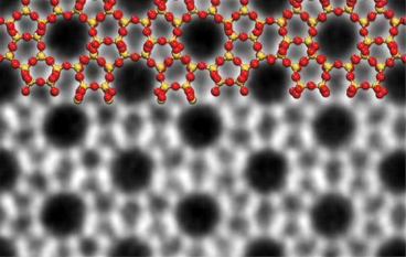
Since zeolite MFI is a widely used catalyst and adsorbent that also holds promise as a thin-film membrane, discovery of nanometre-thick 2D MFI nanosheets has facilitated methods for thin-film zeolite fabrication, which opened new horizons for membrane engineering. In this work, using aberration-corrected scanning transmission electron microscopy, we showed one- to few-unit-cell-wide zeolite MEL intergrowths within 2D-MFI. Our observation of these intergrowths suggests new strategies for the development of ultra-selective zeolite membranes. Read More…

Related Faculty: Andre Mkhoyan

Femtosecond electron imaging of defect-modulated phonon dynamics

electron 8

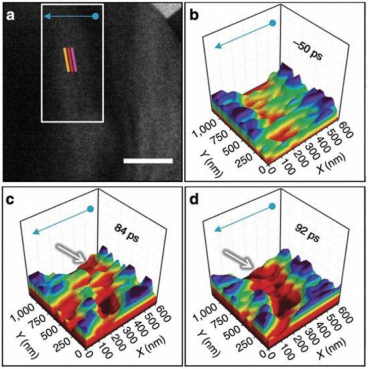
Since precise manipulation and control of coherent lattice oscillations via nanostructuring and phonon-wave interference has the potential to significantly impact the current technologies, resolving the dynamics of individual phonons in defect-laden materials presents an enormous challenge. In this work, we report direct, real-space imaging of the emergence and evolution of acoustic phonons at individual defects in crystalline WSe2 and Ge. Via imaging with an UEM, we were able to image the sub-picosecond nucleation and the launch of wavefronts at step edges and resolve dispersion behaviors during propagation and scattering. These observations provide unique insight into the roles played by individual atomic and nanoscale features on acoustic-phonon dynamics. Read More…

Related Faculty: David Flannigan

High-frequency magnetoacoustic resonance through strain-spin coupling in perpendicular magnetic multilayers

electron 9

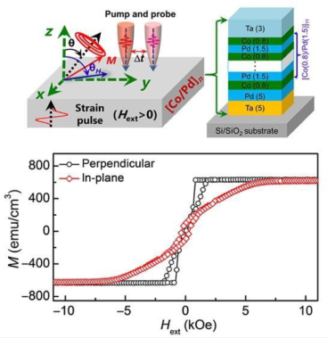
It is desirable to demonstrate an extremely high resonant frequency, assisted by strain-spin coupling, in technologically important perpendicular magnetic materials. In this work, we observe the coupling of magnons and phonons in both time and frequency domains upon femtosecond laser excitation. This strain-spin coupling leads to a magnetoacoustic resonance in perpendicular magnetic multilayers, reaching frequencies in the extremely high band of 60 GHz. We also detail its precise dependence on the magnetostriction. These results offer a potential pathway to manipulating both the magnitude and timing of frequency and strongly coupled magnon-phonon excitations. Read More…

Related Faculty: Xiaojia Wang, Jianping Wang

Metallic line defect in wide-bandgap transparent perovskite BaSnO3

electron 11

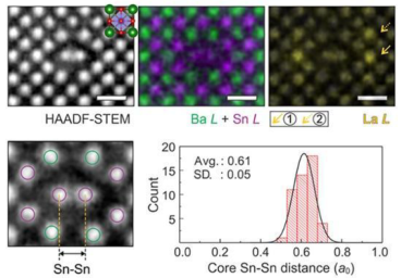
In this work, we report a discovery new line defect with metallic characteristics in optically transparent BaSnO3 perovskite thin films. The distinct atomic structure of the defect core, composed of Sn and O atoms, was visualized by atomic-resolution STEM. The electronic structure of the line defect probed in STEM with electron energy-loss spectroscopy, and supported by ab initio theory, showed the presence of Fermi level–crossing electronic bands that originate from defect core atoms. These metallic line defects act as electron sinks attracting additional negative charges in these wide-bandgap BaSnO3 films. Read More…

Related Faculty: Andre Mkhoyan, Bharat Jalan, Turan Birol

Simultaneous high-strength and deformable nanolaminates with thick biphase interface

electron image

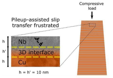
Two-phase nanolaminates are known for their high strength, yet they suffer from loss of ductility. In this paper, we show that broadening heterophase interfaces into 3D interfaces as thick as the individual layers breaks this strength-ductility trade-off. We use micropillar compression and TEM to examine the processes underlying this breakthrough mechanical performance. The 3D interfaces stifle flow instability via shear band formation through their interaction with dislocation pileups. When dislocation pileups fall below a characteristic size relative to the 3D interface thickness, transmission across interfaces becomes significantly frustrated. Read More…

Related Faculty: Nathan Mara

Cryogenic electron microscopy study of nanoemulsion formation from microemulsions

electron 12

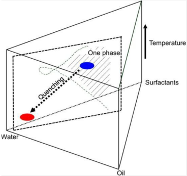
In this work, we examine a process of preparing oil-in-water nanoemulsions by quenching (diluting and cooling) precursor microemulsions made with nonionic surfactants and a cosurfactant. Water-continuous microemulsions produce initial nanoemulsion structures that are small and simple, mostly unilamellar vesicles, but microemulsions that are not water-continuous produce initial nanoemulsion structures that are larger and multilamellar. Examination of these structures by cryo-EM supports the hypothesis that they are initially vesicular structures formed via lamellar intermediate structures, and that if the lamellar structures are too well ordered they fail to produce small simple structures. Read More…

Related Faculty: Alon McCormick, Joseph Zasadzinski